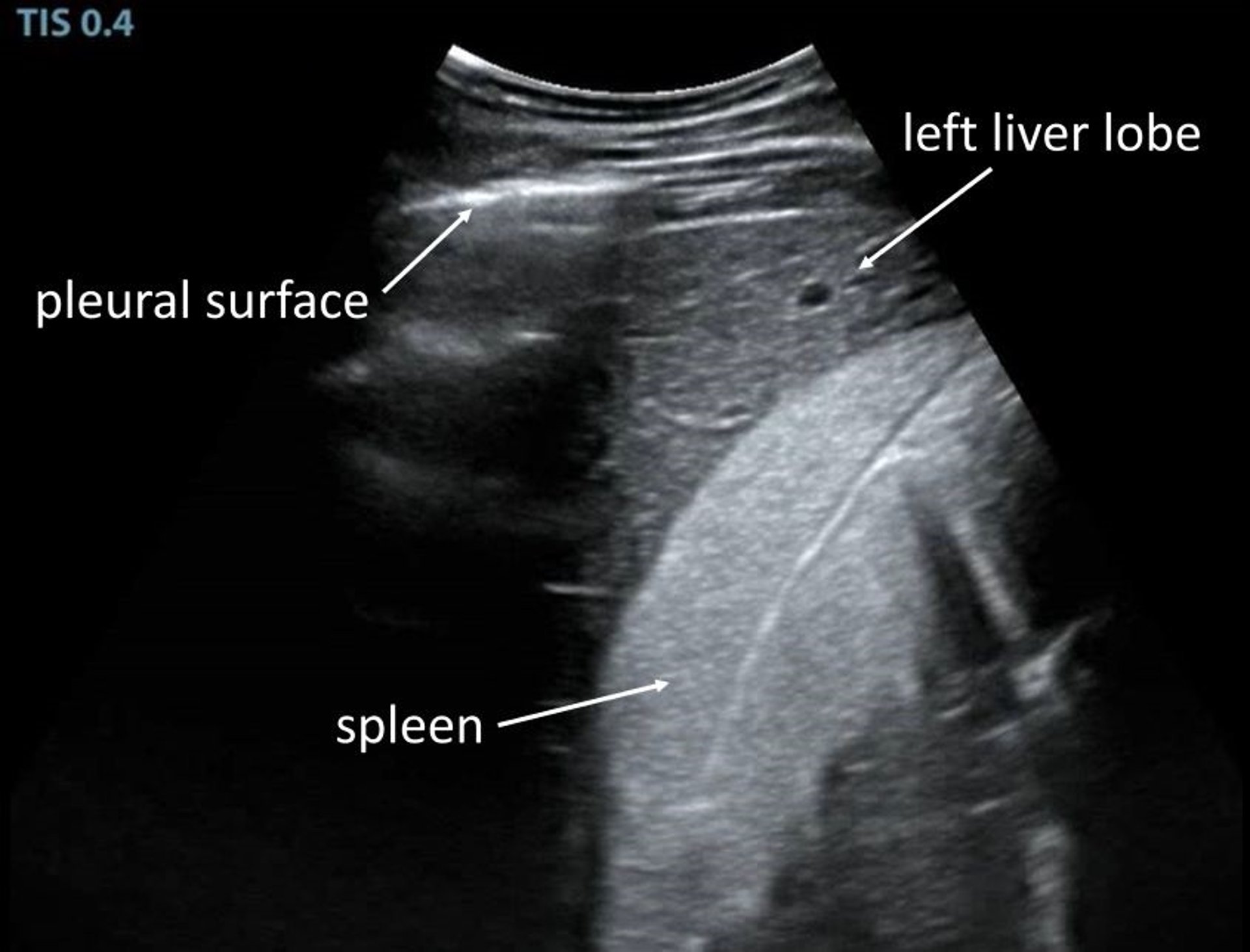
超声图,左肝叶与脾脏,马
Normal ultrasonographic image obtained from the 7th intercostal space in a horse. The left liver lobe can be identified lateral to the spleen, which is slightly more echogenic.
Courtesy of Dr. Amelia Munsterman.
在这些主题中
Normal ultrasonographic image obtained from the 7th intercostal space in a horse. The left liver lobe can be identified lateral to the spleen, which is slightly more echogenic.
Courtesy of Dr. Amelia Munsterman.