P1 骨折,X 光片,马
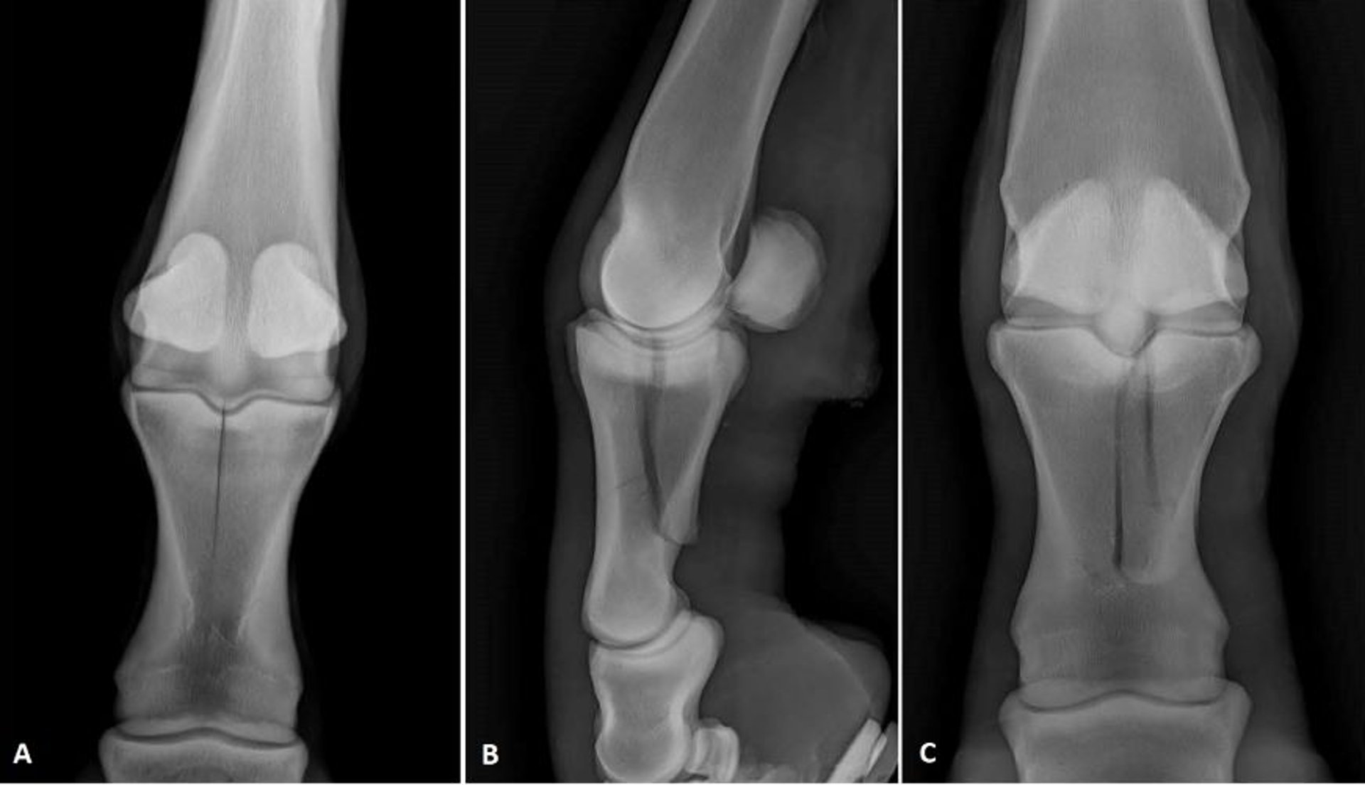
Radiographic images showing two different configurations of proximal phalanx fractures in horses. (A) Dorsopalmar view of a sagittal, incomplete fracture. Lateral (B) and dorsopalmar (C) views of a complicated fracture.
Courtesy of Dr. Matthew T. Brokken.
在这些主题中