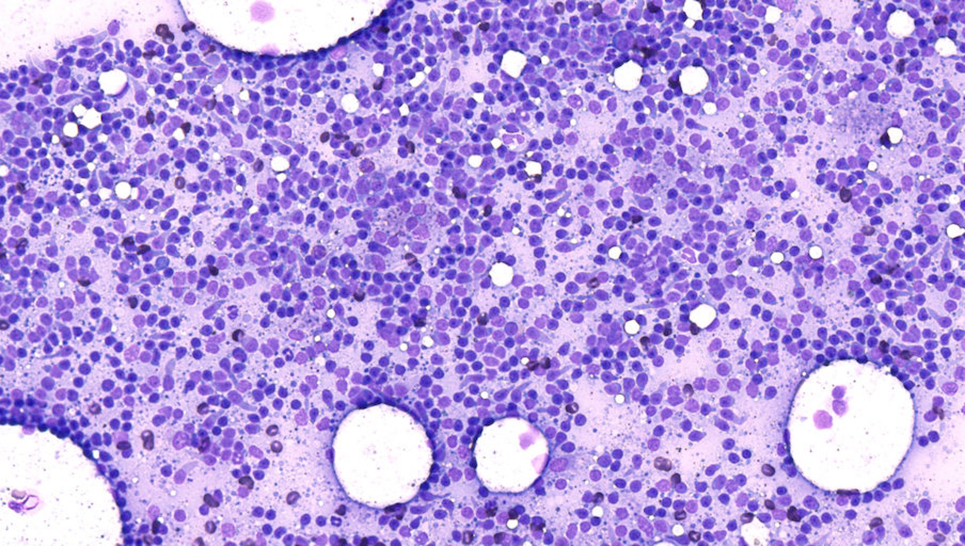
细胞学涂片单层,淋巴细胞
Photomicrograph of an area of the monolayer of a cytologic smear of a lymph node aspirate. The lymphocytes are sufficiently spread to accurately appreciate their size and other morphological characteristics. Approximately half the cells are touching, but no appreciable overlapping is noted. The white circles in the background represent nonstaining lipid material from perinodal adipose tissue. Aqueous Romanowsky stain; original magnification, 200X.
Courtesy of Dr. Rebekah Gunn-Christie.
在这些主题中