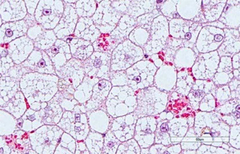
猫肝细胞肿胀显微图像
Photomicrograph of a liver section from a cat with severe microvesicular hepatic lipidosis demonstrating marked hepatocyte distention with triglyceride. Hepatocyte distention has compressed hepatic sinusoids and disturbs perfusion. Note congested RBC in scattered pockets, despite the fact that all hepatocytes abut sinusoidal vasculature. This phenomenon may contribute to transaminase release (ie, ALT and AST) from hepatocytes in cats with severe hepatic lipidosis. H&E stain; scale bar = 60 mcm.
Courtesy of Dr. Sharon Center.
在这些主题中